Research


OUR RESEARCH

Our team works in a multidisciplinary approach using Nanotechnology to combat devastating diseases like Breast and lung cancer. One of the emphases here is overcoming lung cancer mutations induced after using various Tyrosine kinase inhibitors. Our laboratory uses various novel compounds that act as chemosensitizers and help overcome resistance in lung and breast cancers. We are also using exosomes as markers of tumor progression, enabling diagnosis and tumor progression as a liquid biopsy. Another emphasis is to use 3D printing approaches to develop tumor scaffolds and conduct cytotoxicity assays using 3D printed co-cultures of tumor cells and fibroblasts. Apart from this, our team is also printing Cornea using various bioinks.


Mouse model: astrocyte-specific YY1 knockout mice by crossing GFAP-Cre with YY1 loxP mice. This Cre-loxP technology allows us to study the role of YY1 in astrocytes in Mn neurotoxicity.

AREA I: PERCUTANEOUS DELIVERY OF SMALL NOLECULES AND PEPTIDES

Percutaneous delivery provides advantages for skin diseases where the target site is present in the deep epidermis (e.g., fungal, bacterial, viral- infections, psoriasis, dermatitis, and skin cancers like melanoma). In addition, percutaneous delivery offers several advantages over the conventional oral and intravenous dosage forms, such as prevention of the first-pass metabolism, minimization of pain, and possible controlled release of drugs. However, a foremost layer of the skin, the stratum corneum (SC), acts as the main barrier between the body and the environment and limits the delivery of most drugs. To overcome the SC barrier, different nanoparticles such as SLN, NLC, and polymeric nanoparticles were investigated for various small molecules and peptides’ percutaneous delivery.


BILAYERED POLYMERIC NANOPARTICLES FOR COMBINATION THERAPY:

Bilayered polymeric nanoparticles (NPS) are prepared for combination therapy. These NPS are prepared using poly(lactic-co-glycolic acid (PLGA) and chitosan, encapsulating one drug in the PLGA core. In contrast, another drug is encapsulated into a chitosan coat. However, these nanoparticles were unable to reach the deeper skin layers. Therefore the surface of NPS was modified with oleic acid (OA), FDA approved penetration enhancer. During our human skin permeation experiments, we have noted that the surface modification of the NPS with oleic acid (NPS-OA) is responsible for the significant improvement in the spantide (SP) and ketoprofen (KP) delivery to the deeper skin layers.

SP is an anti-inflammatory peptide (MW 1668.76) that antagonizes the neurokinin-1 receptors and thus inhibits the inflammatory response associated with substance-P. KP is a potent non-steroidal anti-inflammatory drug (NSAID) that inhibits arachidonic acid metabolism by potent inhibitory action on cyclooxygenase.

NANOSTRUCTURED LIPID CARRIERS FOR PERCUTANEOUS DRUG DELIVERY:

For percutaneous drug delivery, various lipid carriers such as liposomes solid lipid nanoparticles (SLN) have been well studied. However, liposomes have limitations in their a) physical stability problems with liposomes and b) high costs for effective large-scale production. Similarly, SLNs limit drug expulsion from the carrier and determine drug loading capacity. To overcome this, the second generation of lipid nanoparticles, nanostructured lipid carriers (NLCs), has been developed using the blend of solid lipids and liquid lipids (oils). NLCs have higher loading capacity and lower drug leaching in storage than SLN.

In our efforts to enhance the delivery of NLCs into the deep epidermis, we have already shown that a well-known Cell-penetrating peptide (CPPs), transactivating transcriptional activator (TAT), when linked to nanoparticles, has the potential to carry the payloads across the skin layers. We are screening different CPPs to enhance drug delivery to the deep epidermis. Further, this approach was used to deliver a peptide, spantide II, and small molecule, ketoprofen, together to treat skin diseases like allergic contact dermatitis (ACD).


PH

AREA II: DEVELOPMENT OF ANIMAL MODELS FOR INFLAMMATORY SKIN DISEASES 

Psoriasis Model Imiquimod-induced psoriasis-like skin inflammation in mice.


Allergic Contact Dermatitis (ACD)

Allergic contact dermatitis (ACD) is one of the most prevalent occupational skin diseases. ACD may result from the interaction of xenobiotics with the immune system. ACD is regarded as the emergence of immunotoxicity in humans. Many chemicals have been shown to cause skin sensitization. In common with other forms of allergy, ACD develops in two phases, defined operationally as induction and elicitation. Induction of skin sensitization is initiated by topical exposure of the chemical allergen to an individual sufficient to induce a cutaneous immune response of the necessary vigor, known as sensitization. If the sensitized individual is now exposed subsequently, at the same or a different skin site, to the inducing chemical allergen, then a more vigorous secondary immune response will be provoked at the point of contact. This, in turn, initiates the cutaneous inflammatory reaction known as ACD. This model was sensitized and challenged using 2,4-dinitrofluorobenzene (DNFB).

Psoriasis-like model

Psoriasis is a chronic inflammatory skin disease characterized by erythematous plaques, excessive growth, and aberrant differentiation of keratinocytes, along with increases in angiogenesis and inflammatory cell infiltrate. The most commonly used model for investigating psoriasis is a xenograft model, in which immunodeficient mice are transplanted with human psoriasis-prone skin. However, these experiments are laborious, expensive, and require considerable expertise and technical skills. Therefore, a psoriasis plaque like the model was developed using topical application of imiquimod (IMQ) on the mice’s back skin. This model showed the most features of human psoriasis where cytokines like IL-23 and IL-17 are involved.


PH

AREA III: DELIVERY OF ANTICANCER DRUGS 

This area involves the study of molecular mechanisms involved in anti-cancer therapy using mouse xenograft tumor models. Various lung xenograft models (orthotopic, i.v. administration of cells, s.c. model) are available in the laboratory. The study involves using novel PPAR-gamma agonists against non-small cell lung cancer and understanding their mechanism of action either alone or in combination with Taxotere. Novel approaches for delivering these anticancer drugs are used with a brief outline.


Aerosols

 Formulation of HFA based (134-a and 227) metered-dose inhalers with anticancer drugs, proteins, and Cox-2 inhibitors.

Nebulization of Cox-2 inhibitors to lungs using lung tumor models. A nose-only chamber is available and has been extensively used to evaluate for deposition and efficacy of aerosols in mice.

Formulations of nasal formulations using various polymers and their assessment for droplet size, viscosity, and surface tension using the statistical design of experiments.

Liposomal/Nanoparticle Drug Delivery

Development of various anticancer drugs-antibody conjugates, liposome-containing drugs, liposome-antibody conjugates, and nanoparticle formulations for cancer drug delivery in combination with immunotoxins.

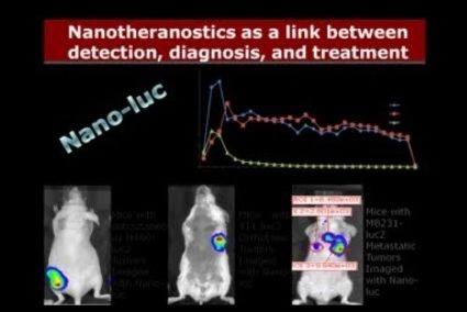
One important part of our research is to enhance the delivery of liposomes to tumors cells by overcoming stromal barriers by using drugs like Telmisartan and Losartan, and withaferin. Apart from this, our research group is also delivering siRNA to tumor cells by using liposomal delivery systems. Imaging of targeted liposomes is also done using various chemoluminescence and bioluminescence markers.

PH  

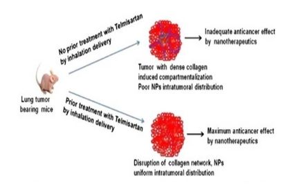
Schematic representation of inhalation use of TEL to reduce tumor fibrosis and increase nanoparticle intratumoral distribution. 

PH  

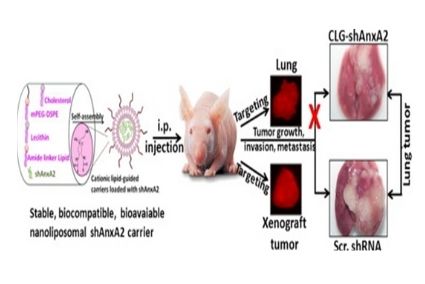
Cationic lipid guided short-hairpin RNA interference of annexin A2 attenuates tumor growth and metastasis in a mouse lung cancer stem cell model.

PH  

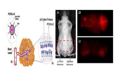
Theranostic tumor homing nanocarriers for the treatment of lung cancer: Nanoparticles conjugated with 6His-PEG2000-CREKA through DOGS-Ni-NTA for targeting tumor vasculature (PCNCs-D)

PH  

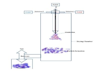
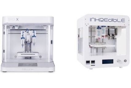
Schematic diagram of the dual-channel spray drying system. Effect of Drug XX on angiogenesis by Tube formation assay.

PH  

In-Vivo Imaging. (A) A549 and H460 lung cancer cell tumor-bering mouse in in-vivo imaging system and Spectrally Unmixed Image of Vasculature with, (B) PCNCs-Di targeting vasculature and (C) NCs-Di. 

PH  

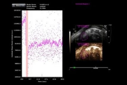
Micro-ultrasound is a real-time modality, molecular imaging, and quantification of angiogenesis using the microbubbles conjugated to ligands targeting VEGFR2 Control tumor-bearing mice. 

PH  


Mouse model: astrocyte-specific YY1 knockout mice by crossing GFAP-Cre with YY1 loxP mice. This Cre-loxP technology allows us to study the role of YY1 in astrocytes in Mn neurotoxicity.

AREA IV: ALTERNATE USE OF ANIMALS USING 3D IN VITRO MODELS

Another component of research in Dr. Sachdeva’s laboratory is the use of various 3D in vitro cultures as an alternative to animals. In this area, two projects are being pursued. The role of 3D cultures as models to study skin irritation has already been demonstrated and various publications are already available in this area.


In vitro 3D culture model for wound healing

Presently there is no wound healing model available in vitro, and most studies are being conducted in vivo. In our laboratory, we are developing a 3D human wound healing model using the EFT-300 cultures from Mattek Corp. Currently, the model is being developed using various corrosive chemicals (e.g., Sulphuric acid, acrylic acid, potassium hydroxide) to induce a wound or heat to induce a burn wound. Various histological changes and the role of various growth factors and cytokines are also being monitored to validate such a model. EFT-300 model consists of epidermis and dermis, which are very important to understand the natural mechanism of wound healing to establish a normal equilibrium of the skin.

In vitro tumor 3D culture model

This project involves the development of an in-vitro tumor model which is more predictive of disease states and drug responses. Algimatrix obtained from Invitrogen is currently being used as the matrix to grow tumor cells as spheroids which will then be treated with various drugs/formulations and perform various mechanistic studies. The objective is to show a correlation between in vitro and in vivo studies, and currently, studies are in progress with lung tumor cells. Algimatrix is pure, non-toxic, has a better nutrient delivery without damage to cells.


Area V

AREA V: SYNTHESIS AND CHARACTERIZATION OF VARIOUS PEG-CHOLECALCIFEROL CONJUGATES FOR OCULAR DRUG DELIVERY

Dry Eye Disease (DED) is an ocular disease caused by the hyperosmolarity of tears manifested by discomfort and visual disturbances, consequently damaging the ocular surface. Topical administration is the most favored route for the treatment of DED. However, frequent drainage from the ocular surface upon administration and poor corneal permeability requires frequent formulations. So there is a need for innovative formulation strategies to overcome these shortcomings. We are designing cholecalciferol PEG conjugates that can function as an ester prodrug for cholecalciferol and self-assemble into nanomicelles due to hydrophilic PEG2000 for the ocular delivery of various drugs such as tacrolimus to treat DED. A mouse model of DED was developed using benzalkonium chloride treatment, and the therapeutic efficacy of cholecalciferol conjugates was evaluated. Progression of DED was monitored by assessing tear pH and corneal staining scores. We observed significant improvement in DED in corneal grading score (1 vs. 4) and reduced inflammatory cells.


CORNEAL PENETRATION STUDY

Fresh porcine eyes were obtained from Bradley Country Store in Tallahassee, FL (slaughterhouse). The anterior portion of the eye was cannulated using a 27 g needle and first flushed and inflated with keratinocyte serum-free medium (KSFM with pituitary extract and EGF) followed by the medium flow rate of 1-2 microlitre/min for the period of study at 37°C. Different formulations were prepared with Coumarin-6 as a tracer dye and applied to eyeballs. Confocal laser scanning microscopy with z-stacking (10μ intervals) was performed. Fluorescence intensities at different depths of the cornea showed significant differences in the different formulations. Nanomicelle gel showed deeper penetration and helped the drug traverse deeper in the corneal layers compared to the nanomicelle solution.

PH  

Corneal fluorescence staining of mouse eye as observed under cobalt blue light using slit microscope (Keeler Inc.).

PH  

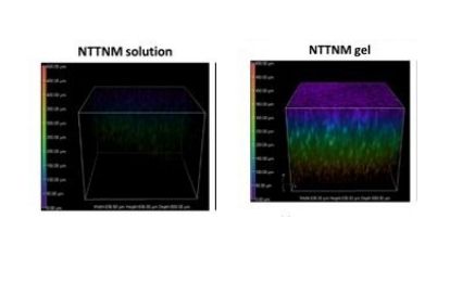
Corneal penetration study in swine eyeballs. Fluorescence with color-coding of depth (z in μm)

PH  

H&E staining of the whole eye (mouse model) (A) KCS control (B) after treatment with Nanoformulation, showing improvement in the thickness of the limbal and central epithelium and stromal regions. 

3D BIO-PRINTING

3D bio-printing technology bridges between the artificially engineered tissue construct with the versatility of native tissue by offering spatial distribution and recapitulation of personalized architectural accuracy. 3D layer by layer printing using vari

FORMULATION AND DEVELOPMENT OF DISSOLVABLE MICRONEEDLES AND TABLETS USING 3D PRINTING TECHNOLOGY

The formulation of dosage forms for the therapeutic delivery of drugs to the systemic circulation is currently seeing advancements in the pharmaceutical industry today. Variations of fabrication techniques are actively being researched, with one of the major players being the additive manufacturing of drugs using 3D printing techniques. In our lab, we are currently working on optimal designs and formulations for both the novel microneedle arrays (MNs) and the traditional tablet drug dosage forms. We have been successful in achieving well-defined 3D-printed MNs. However, we are now working on optimizing the composition of the MNs to demonstrate and advance therapeutic drug dissolution rates in addition to favorable skin permeation rates. Our goal of 3D printing a suitable tablet with favorable dissolution rates to bypass the harsh conditions of the GIT and the liver's tendency to metabolize drugs before reaching the systemic circulation is also coming along with favorable results. Our main driving force in developing this novel form of tablet manufacturing stems from a desire to enable ease of manufacturing and multi-drug tablets. We are using DLP and SLA printing technologies for this purpose and have formulated MNs and tablets by using several model drugs.


FORMULATION 

Biocompatible macromonomeric solutions were purchased from Sigma Aldrich in addition to cytocompatible photoinitiators. The prepolymerized photopolymerizable resin was prepared by varying the amount of macromolecular monomer, photoinitiator and water in the mixture.

3D PRINTING 

VAT polymerization is a unique, highly effective 3D printing technique utilized in this process. High-resolution prints were achieved with the help of our state-of-the-art in-house 3D printers with the ability to print up to 25 micron (X, Y) resolution and 15 micron (Z) resolution. Below are images from our current work as well as previous work in collaboration with the NanoScience Technology Center, the Department of Mechanical and Aerospace Engineering, the Department of Materials Science and Engineering, the Department of Electrical and Computer Engineering, and the Burnett School of Biomedical Sciences, University of Central Florida. The images were obtained from Kundu et al. “DLP 3D Printed “Intelligent” Microneedle Array (iμNA) for Stimuli-Responsive Release of Drugs and Its in-Vitro and ex-Vivo Characterization" Microelectromechanical Systems, Jun. 2020.

PH

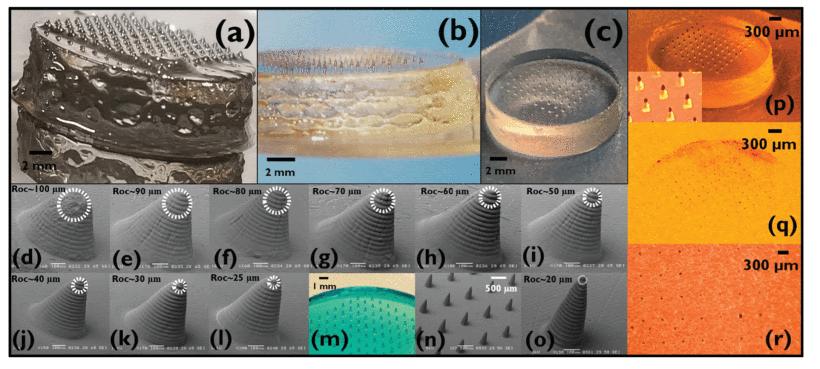
"(a): “Partially Polymerized Membrane” effect on the base on the NA due to background printing from over-exposure, which peels off as the polymer matrix is not completely polymerized in the portions of the structure between the μN bases. (b) “Pancaking” effect on the edges of the iμ NA base. (c) Optimally printed iμ NA. (d-l) Optimization of the aspect ratio (AR) of the iμ NA to successfully achieve the desired ROC to penetrate human skin. (m) iμ NA dyed in methylene blue shows that the polymer matrix retains its hydrogel properties post-printing, allowing for “intelligent” release. (n) SEM image of an optimal aspect ratio iμ NA having a ROC of ∼20 μm (o). (p) Dyeing the tips with Gentian Violet to study the effect of penetration on an artificial skin model. (q) Photomicrographs showing successful penetration of an entire 10×10 array with a close-up image in (r)." Kundu et al. (2020)

PH

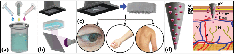
"(a): Schematic of PEGDA (blue) being mixed with 2.5 wt% Diphenyl (2, 4, 6-trimethylbenzoyl) phosphine oxide (TPO) (green) and 0.25wt% therapeutic cargo of diclofenac sodium (pink). (b) DLP 3D printing of the prepared polymer matrix with a UV light source of 385 nm onto the build platform. The iμNA printed consists of a sacrificial base plate and a raft to improve the adhesion of the 3D printed device onto the build platform. (c) Removal of the iμNA from the build platform to have the final iμNA, which may be used for ocular, acute, and chronic drug delivery via transdermal route and allergen testing on the human torso, among other applications. (d) Close-up of a singular API loaded μNA in the intelligent polymer matrix showing the stimulus-based release of the drugs upon sensing its external environment change while retaining the non-drug PEGDA matrix." Kundu et al. (2020)